


Home / Training / Manuals / Atlas of breast cancer early detection / Cases
Atlas of breast cancer early detection
Go back to the list of case studies
.png) Click on the pictures to magnify and display the legends
Click on the pictures to magnify and display the legends
BI-RADS Category (Left BCS, post operative breast): 2 (benign)
| Case number: | 082 |
| Age: | 59 |
| Clinical presentation: | Postmenopausal woman with increased risk of developing breast cancer in view of a history of breast cancer in two of her first-degree relatives, presented with left breast lump. |
Mammography:
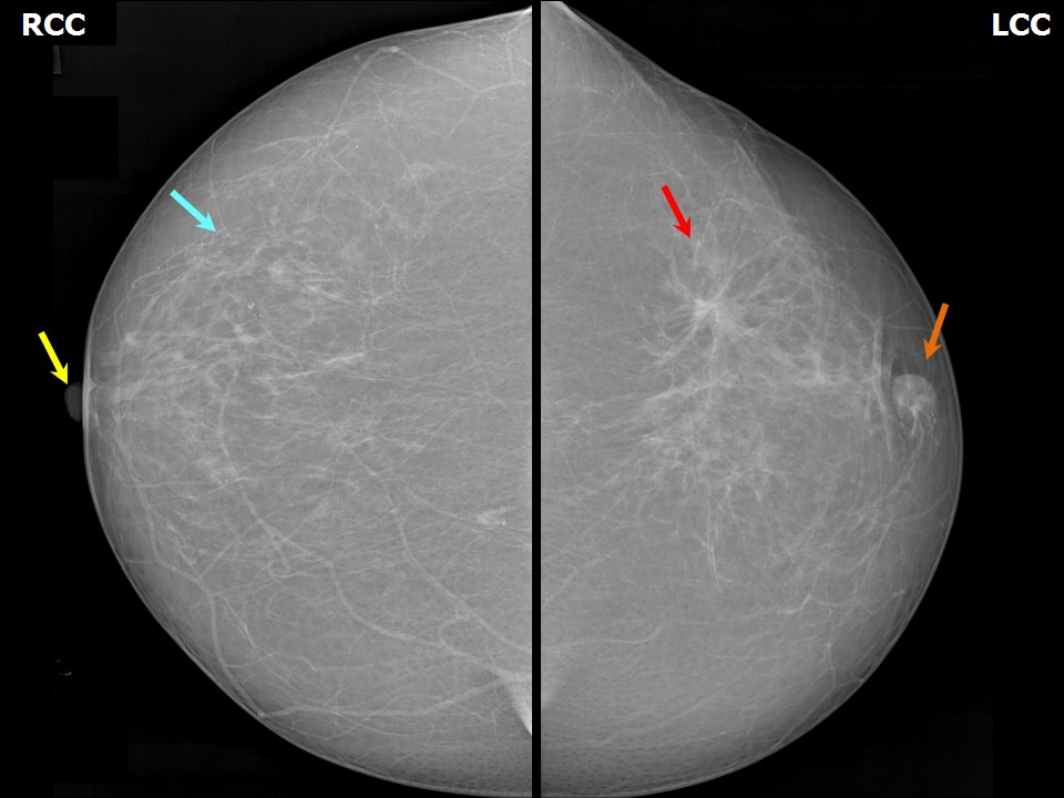 | 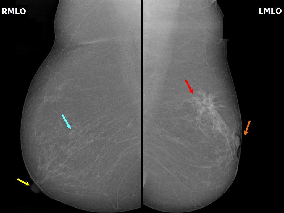 |
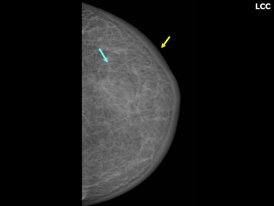
| Breast composition: | ACR category a (the breasts are almost entirely fatty) | Mammography features: |
| ‣ Location of the lesion: | August 2014: Left breast, upper outer quadrant at 12 oclock, anterior and middle thirds |
| ‣ Mass: | |
| • Number: | 1 |
| • Size: | 1.4 × 1.0 cm |
| • Shape: | Irregular |
| • Margins: | Spiculated |
| • Density: | High |
| ‣ Calcifications: | |
| • Typically benign: | None |
| • Suspicious: | None |
| • Distribution: | None |
| ‣ Architectural distortion: | Present |
| ‣ Asymmetry: | Focal |
| ‣ Intramammary node: | None |
| ‣ Skin lesion: | None |
| ‣ Solitary dilated duct: | None |
| ‣ Associated features: | Nipple retraction, axillary adenopathy, and architectural distortion |
 |  |
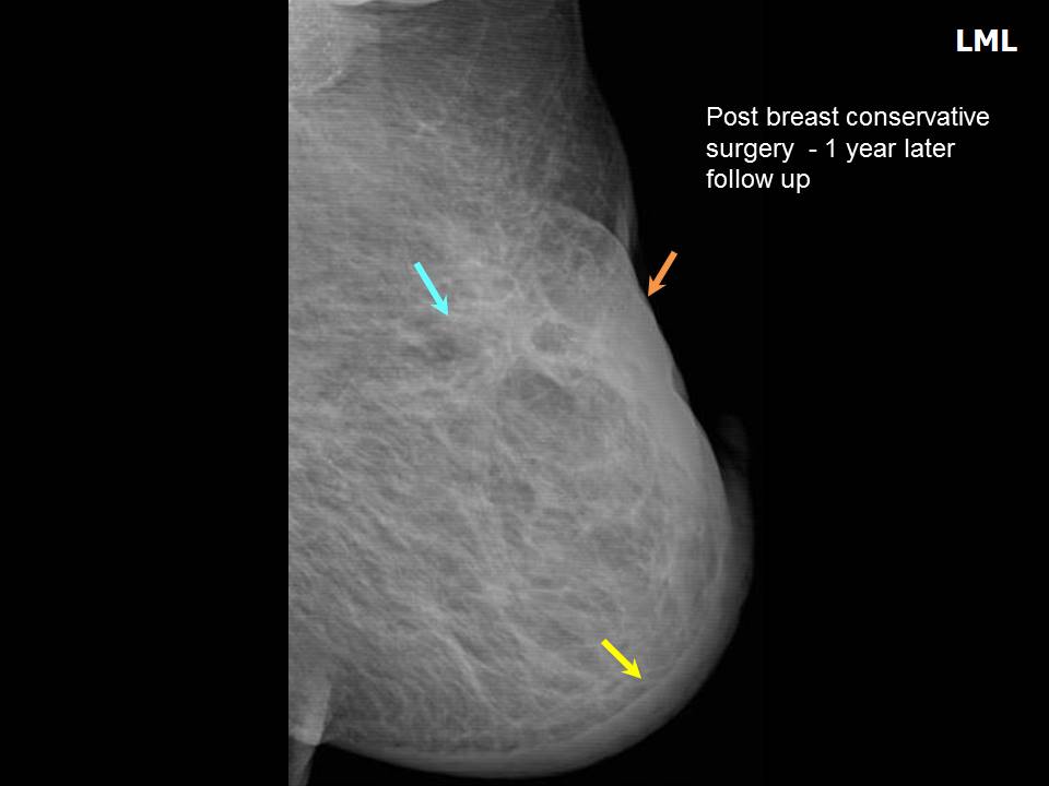 |
| Breast composition: | ACR category b (there are scattered areas of fibroglandular density) | Mammography features: |
| ‣ Location of the lesion: | September 2015: Left breast, upper outer quadrant at 12 oclock, middle third |
| ‣ Mass: | |
| • Number: | 0 |
| • Size: | None |
| • Shape: | None |
| • Margins: | None |
| • Density: | None |
| ‣ Calcifications: | |
| • Typically benign: | None |
| • Suspicious: | None |
| • Distribution: | None |
| ‣ Architectural distortion: | Present |
| ‣ Asymmetry: | Focal |
| ‣ Intramammary node: | None |
| ‣ Skin lesion: | None |
| ‣ Solitary dilated duct: | None |
| ‣ Associated features: | Nipple retraction, skin thickening, trabecular thickening, and architectural distortion |
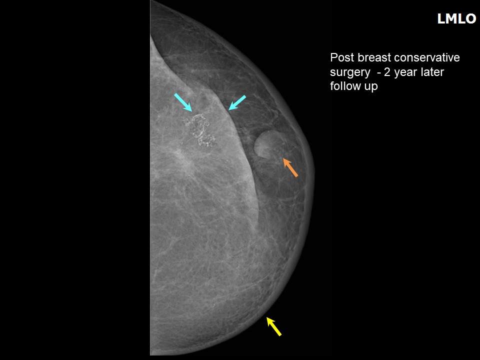 |  |
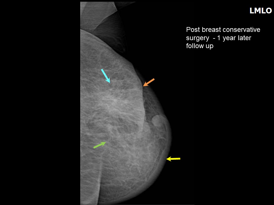
| Breast composition: | ACR category b (there are scattered areas of fibroglandular density) | Mammography features: |
| ‣ Location of the lesion: | October 2016: Left breast, upper outer quadrant at 12 oclock, middle third |
| ‣ Mass: | |
| • Number: | 0 |
| • Size: | None |
| • Shape: | None |
| • Margins: | None |
| • Density: | None |
| ‣ Calcifications: | |
| • Typically benign: | Postoperative dystrophic calcification along surgical scar |
| • Suspicious: | None |
| • Distribution: | None |
| ‣ Architectural distortion: | Present |
| ‣ Asymmetry: | Focal |
| ‣ Intramammary node: | None |
| ‣ Skin lesion: | None |
| ‣ Solitary dilated duct: | None |
| ‣ Associated features: | Nipple retraction, skin thickening, trabecular thickening, and architectural distortion |
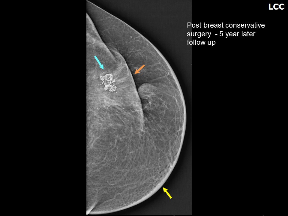 | 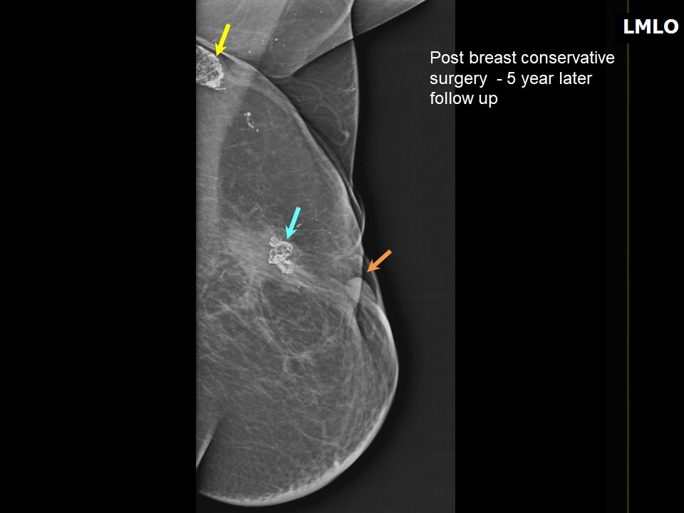 |
| Breast composition: | ACR category a (the breasts are almost entirely fatty) | Mammography features: |
| ‣ Location of the lesion: | November 2018: Left breast, upper outer quadrant at 12 oclock, posterior third |
| ‣ Mass: | |
| • Number: | 0 |
| • Size: | None |
| • Shape: | None |
| • Margins: | None |
| • Density: | None |
| ‣ Calcifications: | |
| • Typically benign: | Postoperative dystrophic calcification along surgical scar in left breast and left axilla |
| • Suspicious: | None |
| • Distribution: | None |
| ‣ Architectural distortion: | Present |
| ‣ Asymmetry: | Focal |
| ‣ Intramammary node: | None |
| ‣ Skin lesion: | None |
| ‣ Solitary dilated duct: | None |
| ‣ Associated features: | Nipple retraction, skin thickening, and architectural distortion |
Ultrasound:
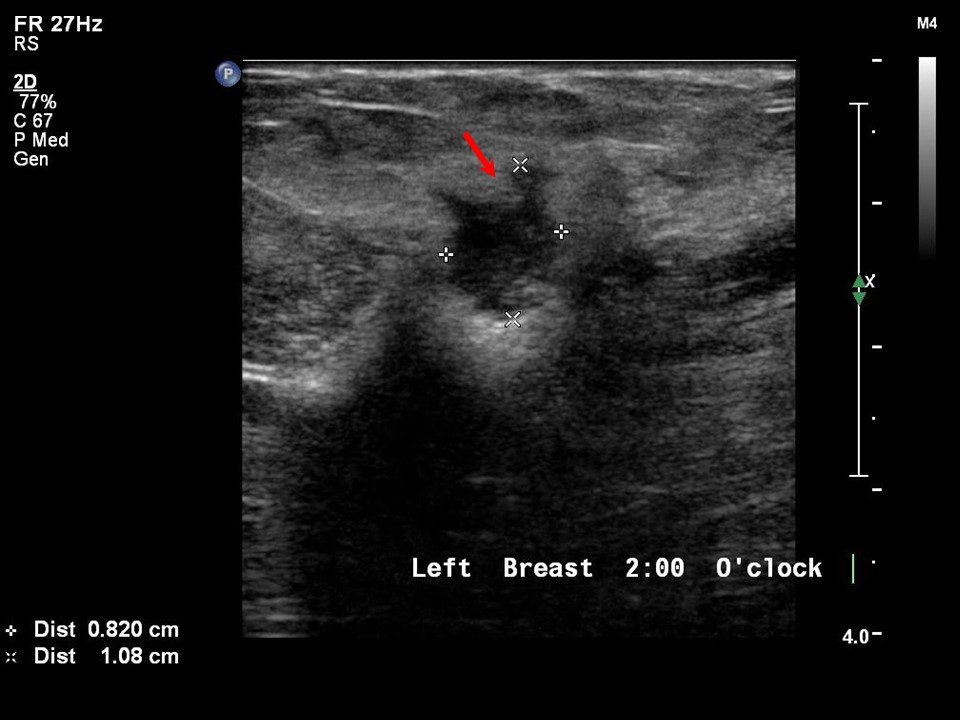 |
| Ultrasound features: August 2014: Left breast, upper outer quadrant at 2 oclock | |
| ‣ Mass | |
| • Location: | August 2014: Left breast, upper outer quadrant at 2 oclock |
| • Number: | 1 |
| • Size: | 1.0 × 0.8 cm |
| • Shape: | Irregular |
| • Orientation: | Not parallel |
| • Margins: | Spiculated |
| • Echo pattern: | Hypoechoic |
| • Posterior features: | No posterior features |
| ‣ Calcifications: | None |
| ‣ Associated features: | Architectural distortion |
| ‣ Special cases: | None |
 |
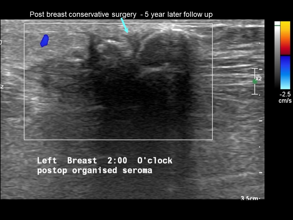
| Ultrasound features: September 2015: Left breast, upper outer quadrant at 2 oclock | |
| ‣ Mass | |
| • Location: | September 2015: Left breast, upper outer quadrant at 2 oclock |
| • Number: | 0 |
| • Size: | None |
| • Shape: | None |
| • Orientation: | None |
| • Margins: | None |
| • Echo pattern: | Heteroechoic |
| • Posterior features: | No posterior features |
| ‣ Calcifications: | None |
| ‣ Associated features: | Architectural distortion along postoperative scar with organized seroma |
| ‣ Special cases: | None |
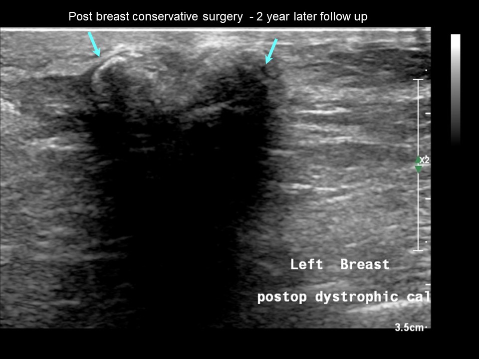 |
| Ultrasound features: October 2016: Left breast, upper outer quadrant at 2 oclock | |
| ‣ Mass | |
| • Location: | October 2016: Left breast, upper outer quadrant at 2 oclock |
| • Number: | 0 |
| • Size: | None |
| • Shape: | None |
| • Orientation: | None |
| • Margins: | None |
| • Echo pattern: | None |
| • Posterior features: | No posterior features |
| ‣ Calcifications: | Macrocalcifications along surgical scar in the organized seroma |
| ‣ Associated features: | Architectural distortion and skin retraction |
| ‣ Special cases: | None |
 |
| Ultrasound features: November 2018: Left breast, upper outer quadrant at 2 oclock | |
| ‣ Mass | |
| • Location: | November 2018: Left breast, upper outer quadrant at 2 oclock |
| • Number: | 0 |
| • Size: | None |
| • Shape: | None |
| • Orientation: | None |
| • Margins: | None |
| • Echo pattern: | None |
| • Posterior features: | No posterior features |
| ‣ Calcifications: | Macrocalcifications along surgical scar in the organized seroma |
| ‣ Associated features: | Architectural distortion, skin retraction, and no vascularity on colour flow mapping |
| ‣ Special cases: | None |
BI-RADS:
BI-RADS Category (August 2014): 5 (highly suggestive of malignancy)BI-RADS Category (Left BCS, post operative breast): 2 (benign)
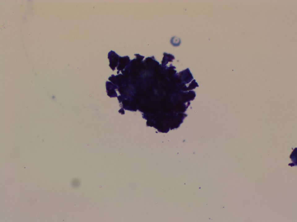
Cytology:
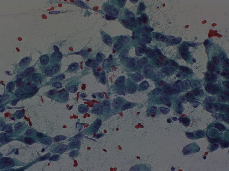 |
| Cytology features: | |
| ‣ Type of sample: | FNAC |
| ‣ Site of biopsy: | |
| • Laterality: | Left |
| • Quadrant: | Upper outer quadrant |
| • Localization technique: | Ultrasound-guided FNAC |
| • Nature of aspirate: | Whitish, blood-tinged |
| ‣ Cytological description: | In 2014: Smears are very cellular, show dyscohesive clusters of pleomorphic malignant cells |
| ‣ Reporting category: | Malignant |
| ‣ Diagnosis: | Carcinoma breast |
| ‣ Comments: | None |
 |
| Cytology features: | |
| ‣ Type of sample: | FNAC |
| ‣ Site of biopsy: | |
| • Laterality: | Left |
| • Quadrant: | Hard lump felt over scar tissue of breast-conserving surgery |
| • Localization technique: | Palpation: Left breast area has a scar measuring 3.5 cm in length. A nodule of 1 cm diameter felt under the scar tissue, hard and gritty feel on needling during FNAC procedure |
| • Nature of aspirate: | Very scant whitish material |
| ‣ Cytological description: | In 2018: Smears from both sites show only basophilic calcific debris. Ductal epithelial cells of breast origin and other cellular material are not seen in the smears |
| ‣ Reporting category: | Insufficient material |
| ‣ Diagnosis: | Negative for malignancy |
| ‣ Comments: | None |
Histopathology:
Breast-conserving surgery
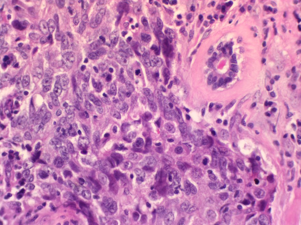 |
| Histopathology features: | |
| ‣ Specimen type: | Breast-conserving surgery |
| ‣ Laterality: | Left |
| ‣ Macroscopy: | Received a lumpectomy specimen oriented with long suture laterally and short suture superiorly. The specimen measures 13 x 10 x 5.0 cm. Skin flap measures 7.0 x 3.5 cm. On serial sectioning a greyish white tumour is identified measuring 4.5 x 4.5 x 2.5 cm. It is located 2.0 cm from the skin, 2.0 cm from the base, 3.0 cm from the superior margin, 3.0 cm from the inferior margin, 3.0 cm from the medial margin, and 2.0 cm from the lateral margin. Whitish firm area on the medial aspect measures 2.0 x 1.5 x 1.0 cm. It is at a distance of 1.0 cm from the tumour. The remaining breast parenchyma is unremarkable. |
| ‣ Histological type: | Invasive breast carcinoma of no special type |
| ‣ Histological grade: | Grade 3 (3 + 3 + 2 = 8) |
| ‣ Mitosis: | 14 |
| ‣ Maximum invasive tumour size: | 7.3 cm (microscopic dimension) |
| ‣ Lymph node status: | 3/10 with extra nodal spread |
| ‣ Peritumoural lymphovascular invasion: | Absent |
| ‣ DCIS/EIC: | DCIS cribriform type, moderate nuclear grade without necrosis |
| ‣ Margins: | Free of tumour However, the invasive carcinoma is seen close to the medial margin, although this margin is technically free of tumour |
| ‣ Pathological stage: | pT3N1 |
| ‣ Biomarkers: | ER positive, PR positive, and HER2 negative |
| ‣ Comments: | There is marked sclerosis with mild lymphocytic response in adjacent breast parenchyma |
Case summary:
| Postmenopausal woman with increased risk of developing breast cancer presented with left breast lump. Mammography and breast ultrasound revealed irregular mass lesion in left breast. Diagnosed as highly suggestive of malignancy, BI-RADS 5 on imaging, as left breast carcinoma, high grade on cytology, and as invasive breast carcinoma, PT3N1 on histopathology. On postoperative surveillance, she presented with a hard lump and pain at the site of surgery in the left breast. Now diagnosed as left breast postoperative organized seroma with dystrophic calcification, BI-RADS 2. |
Learning points:
|